Dr. Aya Ben Yakov
How is real-life experience transformed into discrete memories, and how are those memories stored and retrieved? We aim to answer these questions using a range of naturalistic stimuli (movies, stopmotion, and animation), combining behavioral and brain imaging methods to illuminate the underpinnings of memory formation and organization in healthy individuals.
For more information, please visit the lab’s website.
Dr. Gadi Gilam
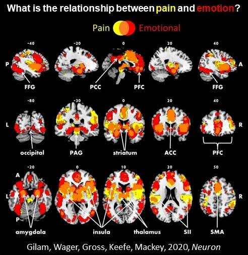
The translational Social Cognitive and Affective Neuroscience (tSCAN) lab investigates mechanisms underlying and influencing the relationship between pain and emotions, as they manifest at the intersection of chronic pain (e.g. Fibromyalgia, Migraine) and psychopathological (e.g., Depression, Anxiety) conditions. Current projects focus on interoception, injustice, emotion regulation, social isolation vs. interactions, usage of body maps, and the link between pain and trauma. To address these issues, the tSCAN lab uses a bio-psycho-social approach, combining cognitive neuroscience, experimental psychology, and health informatics methods, while integrating perspectives from emotion science, social and health psychology, pain medicine, and psychiatry, among others.
For more information, please visit the lab’s website.
Dr. Yuval Hart
We are interested in the neural mechanism underlying creative search. Our lab developed a novel paradigm, the Creative Foraging Game, that measures the process of creative search during an online game of creating shapes out of ten squares. Participants move one square at a time to create beautiful and interesting shapes. The task provides many measures of the search process – exploration and exploitation bouts, originality, efficiency of the search, fluency, flexibility, and many more. We scan subjects in resting-state and use qMRI to characterize their connectivity patterns as well as scan their activity and connectivity patterns during the game.
For more information, please visit the lab’s website.
Dr. Shir Atzil
Why do humans invest substantial energy in socializing, and is there a unifying benefit across different bonds? Social interactions demand significant cognitive and metabolic resources, suggesting they provide immediate biological rewards. We propose that social interactions reduce the metabolic costs of physiological regulation, offering a Social Physiological Gain. We term this phenomenon Social Physiology and propose that it incentivizes social behavior and shapes partner preferences based on physiological benefits.
In this research, we empirically test the concept of Social Physiology by examining glucose and stress metabolism to map the neurobiological pathways—from brain to body—through which a romantic partner influences physiology, revealing the brain-body axes of control modulated by social bonding.
For more information, please visit the lab’s website.
Prof. Shahar Arzy
We are interested in exploring the (human) experiencing-self and its relations to the surrounding environment: the space around us, the events that make up our life and future plans, the people we encounter along the way, and the concepts we develop throughout. We explore the psychological, computational, and neural mechanisms supporting these relations and their role in neuropsychiatric disorders, with a specific focus on Alzheimer’s disease. We hope to generate novel theoretical, computational, and neural understandings of the mechanisms involved in these orientation processes as well as design useful tools to help people and patients orient and re-orient themselves in our dynamic world.
For more information, please visit the lab’s website.
Prof. Aviv Mezer
- Multiple sclerosis and white matter disorders:
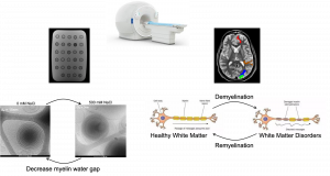
We develop a quantitative MRI approach for the non-invasive mapping of the myelination biomarker revealed by the myelin water gap. In postmortem studies, it was shown that the water layer gap between healthy myelin membranes is compact. However, it changes during the demyelination and remyelination processes. We developed a biophysical model to estimate the water gap, designed an in vitro lipid system with varying water gaps, and validated it using cryo-TEM. Our model successfully estimates the water gap in vitro and shows a reliable estimation for in vivo
- Parkinson:
Parkinson’s disease (PD) is a progressive neurodegenerative disorder dominated by motor and non-motor dysfunction. Despite extensive research, the in vivo characterization of PD-related microstructural brain changes remains an ongoing challenge, limiting advancements in diagnostic and therapeutic strategies. In this study, we collected multiparametric quantitative MRI (qMRI) brain data of PD patients and healthy controls, to investigate microstructural alterations in the PD subjects and in healthy aging. We utilized multiple techniques to analyze the spatial variations of various qMRI parameters, including relaxation rates (R1, R2, R2*), water fraction (WF), susceptibility, magnetization transfer saturation (MTsat), and diffusion metrics (MD, FA). The new qMRI dataset provides valuable insights into PD pathology and healthy aging.
For more information, please visit the lab’s website.