Unit Facilities
Confocal microscope - Leica Stellaris 5 with a super-continuum white light laser
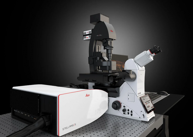
Key features:
- Fluorescence excitation at any arbitrary wavelength in the 485-685nm range, besides the conventional 405nm DAPI excitation channel.
- Separating spectrally-overlapping fluorophores based on their fluorescence lifetime, and user-selectable collection wavelength ranges.
- Built-in algorithms for image deconvolution and automated analysis.
- A confocal reflectance mode that can be used for label-free imaging of axonal myelination.
- Best in-class user friendliness.
Wide-field fluorescence microscope - Olympus IX83P2ZF
This microscope is designed for fast, sensitive imaging of large sections.
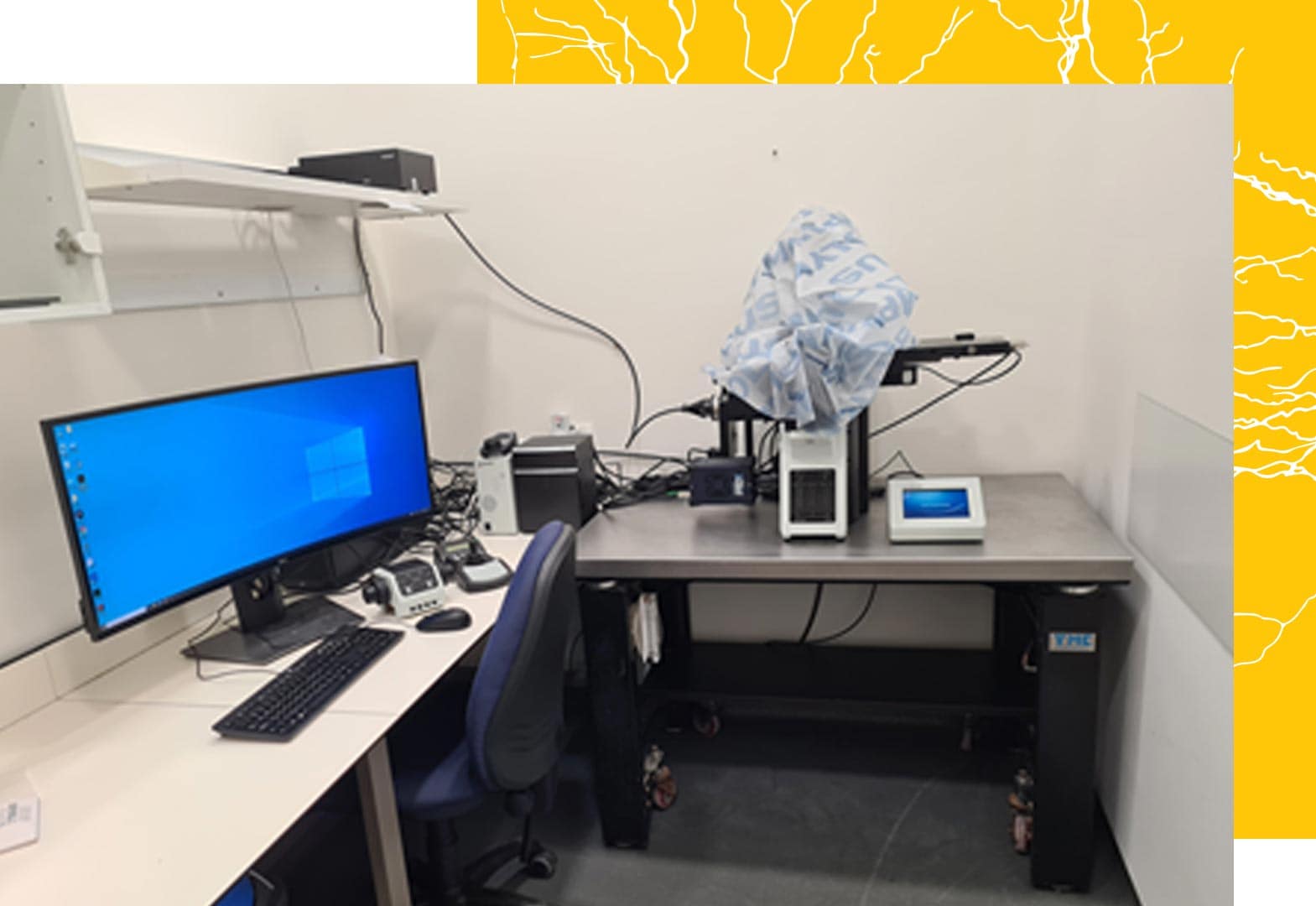
Key features:
- 9-slide stage
- Plate stage
- Universal dish stage (35mm and 60mm)
- 95% Quantum efficiency camera: 2048×2048 pixels with 16-bit depth
- 6 separate illumination LEDs, 7 excitation filters, 6 dichroic mirrors and 7 emission filters for every practical combination of excitation and emission.
- Fast filter wheels
- A wide variety of objective lenses:
- 4x
- 10x
- 20x – long working distance
- 20x – high NA
- 40x
- 60x water
- Hardware-based, fast, continuous autofocus (not an option for 4x magnification).
- Quad dichroic mirror for high-speed channel switching (DAPI, GFP, mCherry, Cy5) and individual mirrors with high sensitivity (for CFP, YFP, GFP, mCherry, Cy5).
LED excitation lights – Lumencor spectraX
- Violet: 395/25nm, 295mW
- Blue: 440/20nm, 256mW
- Cyan: 470/24nm, 196mW
- Teal: 510/62nm, 62mW
- Green-Yellow: 570/80nm, ~600mW
- Red: 640/30nm, 231mW
Excitation filters (on fast wheel):
- ET395/25x
- ET470/40x
- ET485/25x
- ET550/20x
- ET560/40x
- ET620/60x
- ET640/30x
- Free
ET436/20x – permanent on blue LED
Dichroic mirrors (separate wheel):
- 89100bs – sedat quad for fast switching
- T455lp
- T495lpxr
- T565lpxr
- T585lpxr
- T660lpxr
- Open slot for custom filter cubes
- Free
Emission filters (fast wheel):
- ET455/50 nm
- ET480/40 nm
- ET525/36 nm
- ET525/50 nm
- ET605/52 nm
- ET630/75 nm
- ET705/72 nm
- Free
To identify the right filter/fluorophore combination for your experiment, please use: https://www.fpbase.org/spectra/ This tool supports all catalog numbers for components and all possible fluorophores.
Multi-photon microscopes - LOTOS
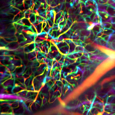
These two microscopes are designed for deep imaging through light-scattering tissues, such as the living brain.
Key features:
- Two independent microscopes (Cajal & Golgi), both fed by a Chameleon Vision II femtosecond laser.
- Acquisition rates of up to 1,000 frames per second using resonant-galvo scanners, implementing Low-power Temporal Over-Sampling (LOTOS) imaging for reduced photodamage.
- PMT2101/M GaAsP PMTs.
- Rapid beam power modulation using Conoptics 350-105 / 350-50 electro-optic modulators and 302RM high voltage amplifiers.
- Light-tight, soundproof Faraday cages.
- Ultra-stable beam alignment using 3D Optix optomechanics.
Future upgrades:
- Time-tagged photon counting with a temporal precision of 42 picoseconds, for improved signal-to-background and ΔF/F ratios.
- Fluorescence lifetime microscopy, second harmonic generation (SHG) and gated imaging.
- Rapid volumetric imagery of the living brain at up to 320 volumes per second. (Pending user demand)
- Super-continuum femtosecond excitation for deeper, non-degenerate two-photon microscopy at ~1,300nm and ~1,650nm. (Pending user demand)
Light Sheet Microscope - La-Vision Ultramicroscope II
This microscope is designed for fast, high-resolution scans of large cleared samples (e.g., with iDISCO or Clarity).
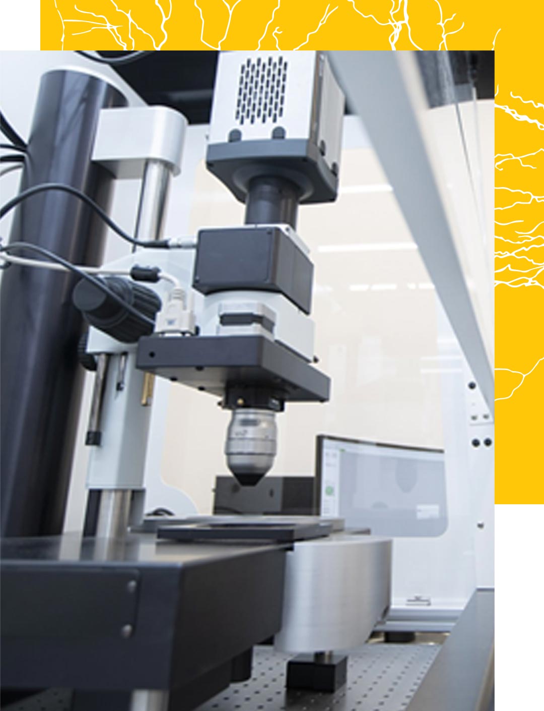
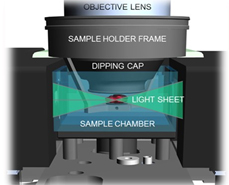
Key features:
- A chemical hood for safe work with organic solvents (e.g., DBE and ECi).
- 3 sheets for minimal light obstruction.
- A huge working distance (> 9mm) for most objective lenses
- High-end sCMOS camera: 2,560×2,160 pixels, pixel size 6.5×6.5 μm²
- Dynamic focus for axial sweeping.
- Fixed lens configuration:
- 3x/2.6x
- 4x/8x
- 12x/24x
- Zoom body configuration
- 63-6.3x adjustable zoom
Excitation lasers:
- 405nm: 100mW
- 488nm: 85mW
- 561nm: 100mW
- 639nm: 70mW
- 785nm: 75mW
Emission filters:
- 460/40 nm
- 525/50 nm
- 620/60 nm
- 680/30 nm
HIVE analysis server
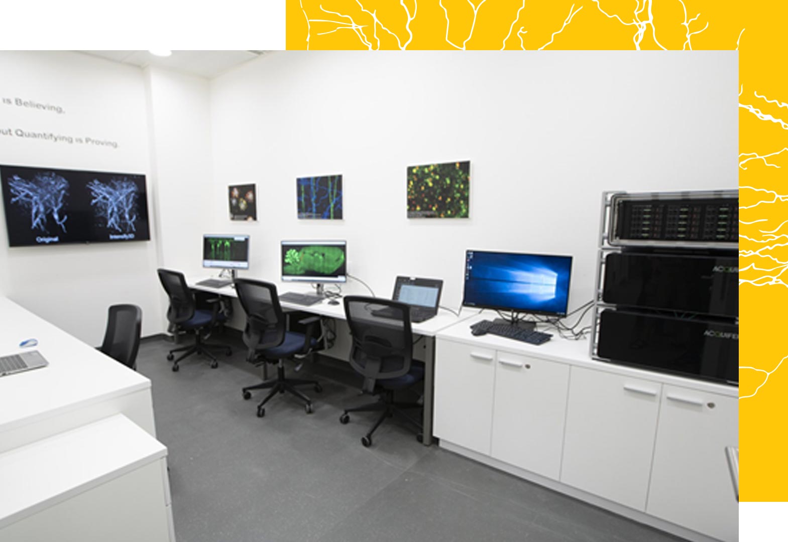
The HIVE is an analysis and communication server designed for imaging core facilities. It is composed of three servers:
- A network server, which is connected directly by 10 Gbit wiring to each microscope and allows data transfer rates of up to 1 Gbit per second, facilitating online analysis and direct acquisition to the HIVE storage. In addition, the server provides fast remote access to the HIVE with a personalized Windows environment.
- A storage NAS RAID6 server, which supplies 52TB (easily expandable) of super high-speed (up to 2.2 Gbit per second) safe storage for analysis.
- A core Windows 2019 processing server, which holds high-speed 512 GB RAM (expandable to 2 TB) with Intel Xeon Silver 20 Core/40 thread CPUs and NVIDIA RTX8000 with 48 GB GPU-RAM (biggest capacity GPU on the market).
For more information, see: https://www.acquifer.de/data-solutions/
Surgery room
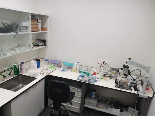
Diagnostic equipment and inventory
