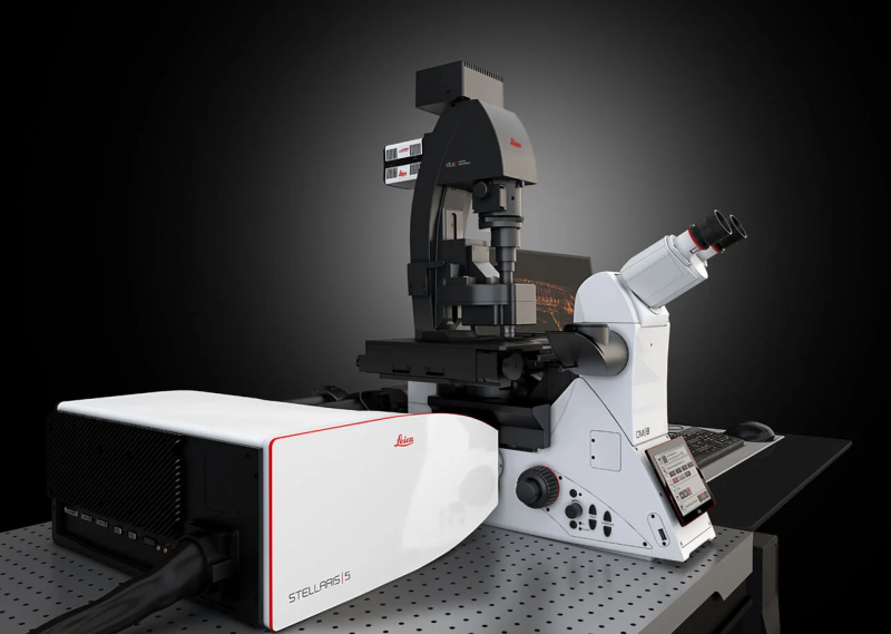
Key features:
- Fluorescence excitation at any arbitrary wavelength in the 485-685nm range, besides the conventional 405nm DAPI excitation channel.
- Separating spectrally-overlapping fluorophores based on their fluorescence lifetime, and user-selectable collection wavelength ranges.
- Built-in algorithms for image deconvolution and automated analysis.
- A confocal reflectance mode that can be used for label-free imaging of axonal myelination.
- Best in-class user friendliness.
Wide-field fluorescence microscope – Olympus IX83P2ZF
Key features:
- 9-slide stage
- Plate stage
- Universal dish stage (35mm and 60mm)
- 95% Quantum efficiency camera: 2048×2048 pixels with 16-bit depth
- 6 separate illumination LEDs, 7 excitation filters, 6 dichroic mirrors and 7 emission filters for every practical combination of excitation and emission.
- Fast filter wheels
- A wide variety of objective lenses:
- 4x
- 10x
- 20x – long working distance
- 20x – high NA
- 40x
- 60x water
- Hardware-based, fast, continuous autofocus (not an option for 4x magnification).
- Quad dichroic mirror for high-speed channel switching (DAPI, GFP, mCherry, Cy5) and individual mirrors with high sensitivity (for CFP, YFP, GFP, mCherry, Cy5).
To identify the right filter/fluorophore combination for your experiment, please use: https://www.fpbase.org/spectra/ This tool supports all catalog numbers for components and all possible fluorophores.
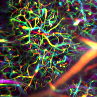
These two microscopes are designed for deep imaging through light-scattering tissues, such as the living brain.
Key features:
- Two independent microscopes (Cajal & Golgi), both fed by a Chameleon Vision II femtosecond laser.
- Acquisition rates of up to 1,000 frames per second using resonant-galvo scanners, implementing Low-power Temporal Over-Sampling (LOTOS) imaging for reduced photodamage.
- PMT2101/M GaAsP PMTs.
- Rapid beam power modulation using Conoptics 350-105 / 350-50 electro-optic modulators and 302RM high voltage amplifiers.
- Light-tight, soundproof Faraday cages.
- Ultra-stable beam alignment using 3D Optix optomechanics.
Future upgrades:
- Time-tagged photon counting with a temporal precision of 42 picoseconds, for improved signal-to-background and ΔF/F ratios.
- Fluorescence lifetime microscopy, second harmonic generation (SHG) and gated imaging.
- Rapid volumetric imagery of the living brain at up to 320 volumes per second. (Pending user demand)
- Super-continuum femtosecond excitation for deeper, non-degenerate two-photon microscopy at ~1,300nm and ~1,650nm. (Pending user demand)
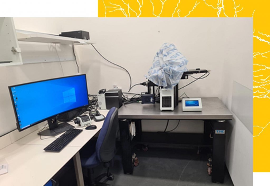
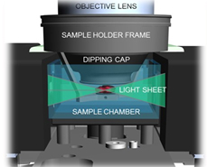
This microscope is designed for fast, high-resolution scans of large cleared samples (e.g., with iDISCO or Clarity).
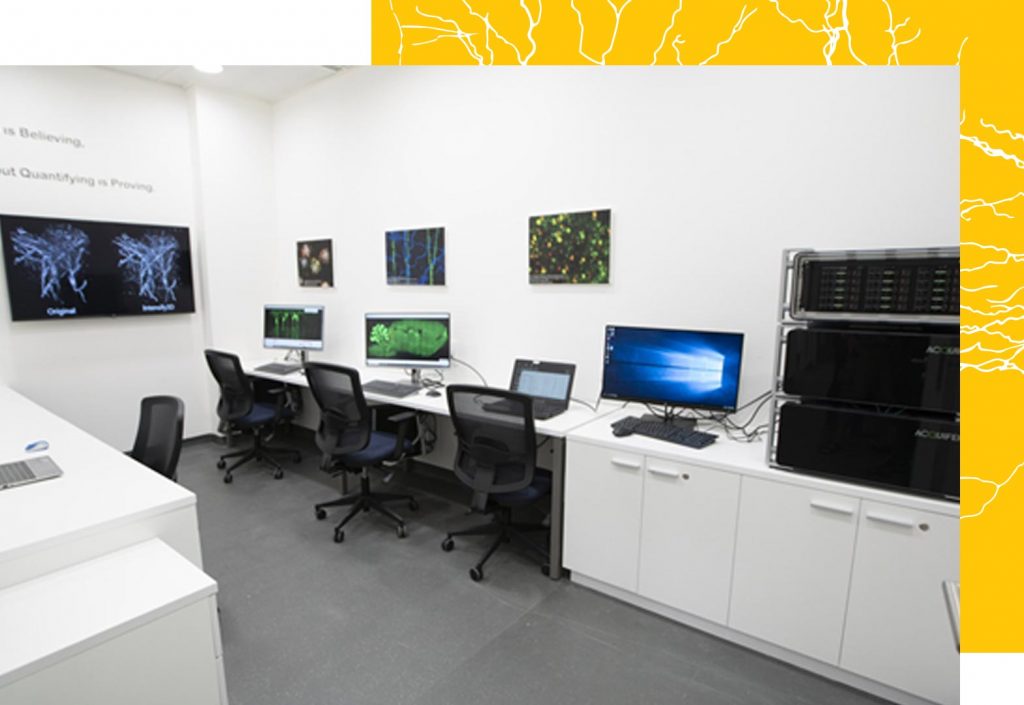
The HIVE is an analysis and communication server designed for imaging core facilities. It is composed of three servers:
- A network server, which is connected directly by 10 Gbit wiring to each microscope and allows data transfer rates of up to 1 Gbit per second, facilitating online analysis and direct acquisition to the HIVE storage. In addition, the server provides fast remote access to the HIVE with a personalized Windows environment.
- A storage NAS RAID6 server, which supplies 52TB (easily expandable) of super high-speed (up to 2.2 Gbit per second) safe storage for analysis.
- A core Windows 2019 processing server, which holds high-speed 512 GB RAM (expandable to 2 TB) with Intel Xeon Silver 20 Core/40 thread CPUs and NVIDIA RTX8000 with 48 GB GPU-RAM (biggest capacity GPU on the market).
For more information, see: https://www.acquifer.de/data-solutions/
The surgery room allows you to prepare your animals or fixed samples for imaging at one of our multi-photon microscopes. Among others it is equipped with a sevoflurane evaporator for anaesthesia induction, a stereomicroscope, a stereotaxic frame, a dental drill, a regulated heat pad, a freezer and a fridge
Our diagnostic instruments are aimed at quickly identifying and resolving issues you’re facing with your experimental equipment. Our inventory of optical, mechanical and electrical parts is meant to let you set up your experiment quickly, without having to wait for some parts to be shipped across the globe. It contains hundreds of different parts. Please let us know if additional parts could assist your research.