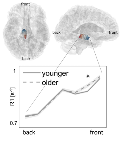
Our article presents a method for estimating the change in MRI measurements along the shape of a given brain structure. Here, we focus on the midbrain (Figure 1), which contains numerous nuclei and white-matter tracts. These midbrain structures are involved in motor, auditory, and visual processing, and show morphological alterations that are associated with aging and with neurological diseases.
We chose to calculate structural profiles along the midbrain by relying on morphology: First, we estimated the midbrain’s main spatial axes (the first axis can be seen in Figure 1); then, along these axes we sampled quantitative MRI measurements that are sensitive to the brain tissue’s biological properties, such as iron and water concentration. To show that these profiles are sensitive to the midbrain’s underlying macrostructure, we compared the profiles to the midbrain’s subregions. We found that the midbrain’s microstructure profiles are sensitive to the age of the subjects. For example, Figure 1 shows that in the frontal segment of the midbrain, older subjects have increased levels of R1, an MRI measurement. This increase is specific to this segment of the midbrain, which overlaps mainly with the substantia nigra (a region that is involved in Parkinson’s disease). This finding highlights the advantage in this sampling approach when studying midbrain structure. Our results were stable across image resolutions and datasets, further supporting the theory that the profiles reflect the shared properties of the midbrain’s micro- and macrostructure.
Figure 1:

