When we are motivated, we tend to do better. For example, when athletes are playing for high gains during crucial matches they often step up their game. In the lab, we study this phenomenon by manipulating the potential rewards animals may receive upon successfully completing a task. In this paper, we investigated how the expectation for future reward affects the activity of neurons in the brain. We focused on two major motor areas of the brain: the basal ganglia and the cerebellum. In the past, neural reward signals were thought to be confined mainly to the basal ganglia. However, recent research from our lab and others has shown that reward signals appear in the cerebellum as well. This was a surprising discovery, as previously the cerebellum had been understood to be a primarily motor area. To relate reward signals in these two areas, and gain a better understanding of the way reward expectations influence the brain as a whole, we collected an extensive and unique dataset of recording of neural activity from these two brain structures simultaneously in the same monkeys.
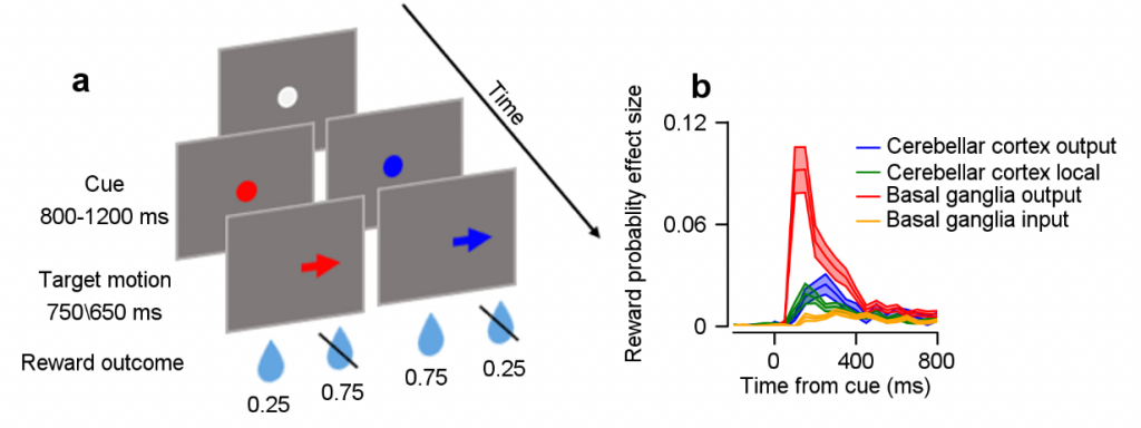
In this dataset, we collected neural recordings from the basal ganglia and cerebellum of two monkeys as they performed an eye-movement task under different motivation conditions. Each trial in our task began with the monkey fixating on a colored dot in the middle of a screen (Fig. a). The monkey associated the color of the spot with the probability of receiving a food reward at the end of the trial (for example, a red dot yielded a reward 25% of the time, while a blue dot yielded a reward 75% of the time). Then the spot moved to one of eight possible locations on the screen and the monkey had to track it with its eyes.
This allowed us to investigate the neurons’ response to change in the color of the target, that is, the time in which reward information is first made available to the monkey. Our sample contained an output population of the basal ganglia and cerebellar cortex, and an input or local population within each structure. Fig. b shows a quantification of the degree to which the activity of neurons in cerebellum subpopulations (blue, green) and basal ganglia subpopulations (red, yellow) was informative as to the reward expectation condition (b). We found that reward expectation signals in a specific subpopulation of the cerebellum are larger than in populations that are canonically associated with reward processing (comparison between the yellow trace and the green and blue ones in b). Moreover, we found that while reward signals were increased between input and output populations in the basal ganglia (yellow versus red), this was not the case in the cerebellum (blue versus green), implying that these signals are processed differently within the structures.