This research project arises from an important discovery of Prof’ Dror Sharon from the Department of Ophthalmology of the Hadassah Medical Center with whom I collaborate. He discovered that individuals with specific mutations in one particular gene (DHDDS) suffer a disease (Retinitis Pigmentosa) that gradually leads to blindness. Usually, individuals with this disease show mutations in Rodohopsin (essential for vision) and normal DHDDS protein, which does not serve the seeing process. However, in the newly discovered disease the rhodopsin gene is normal and only the DHDDS gene has mutations. Importantly, unlike the Rhodopsin protein that appears only in the retina the DHDDS protein appears in virtually every cell of the body.
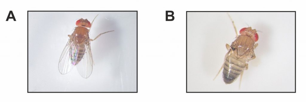
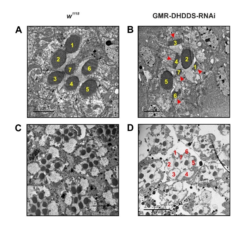
How malfunction of a ubiquitously expressed enzyme leads to specific retinal disease was a great puzzle that could not be addressed in human patients. Therefore, we undertook the challenge of developing an animal model suitable for investigating the cellular and molecular mechanisms responsible for this retinal disease. To this end, we exploited the fact that DHDDS is highly conserved through evolution and can be found in all animal species, including the fruit fly Drosophila. Accordingly, we used the molecular genetic power of Drosophila, which permits the suppression of expression of DHDDS in any targeted tissue. Figure 1 wing loss, and figure 2 shows that targeted knockdown of DHDDS in the developing retina results in a unique pattern of retinal degeneration.
Importantly, we found that the photoreceptor cells in this retina are abnormal and show a drastic reduction of rhodopsin concentration. These phenomena are known to result in photoreceptors degeneration. Our paper describes the mechanisms by which DHDDS cause this degeneration.

Fig. 1. Knockdown of DHDDS in the developing wing resulted in a complete wing loss.
Panels A, B show representative photographs, taken under a stereo-microscope, of the wings of F1 progenies, A, Control flies. B, DHDDS-RNAi flies (mutants)showing wing absence, indicating that DHDDS is essential for wing formation.

Fig. 2. Retinal transmission electron microscopy (TEM) sections of DHDDS-RNAi knockdown flies revealed a unique pattern of photoreceptors degeneration.
Thin TEM sections of control retina (A, in high magnification, C, in lower magnification) and knockdown of DHDDS in theretina (B, D). The identity of the various photoreceptors in each eyelet (ommatidium) is indicated by numbers.


