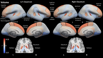
The claustrum is a thin sheet of neurons deep within the white matter. It is highly interconnected with sensory, frontal, and subcortical regions, yet its function is still largely unknown. The deep location of the claustrum, with its fine structure, have limited the degree to which it could be studied in vivo. Particularly in humans, identifying the claustrum using MRI is extremely challenging, even manually. Therefore, automatic segmentation of the claustrum is an invaluable step toward enabling extensive and reproducible research of the anatomy and function of the human claustrum. In this study we developed an automatic algorithm for segmenting the human claustrum in vivo using high-resolution MRI. Using this algorithm, we segmented the claustrum bilaterally in 1,068 subjects of the Human Connectome Project (HCP) Young Adult dataset. We used the resulting segmentations to perform a structural covariance analysis and uncover the brain network of regions that covary with the claustrum. The results show the high connectivity of the claustrum with various brain regions. The automatic segmentation could be valuable for future studies addressing the function of the claustrum.
“Structural correlation results for region volume. The top 12 most correlated regions (in absolute value) in terms of volume are shown for the left and right claustra. Each brain region is colored according to the correlation with the claustrum. Cortical regions are presented on FreeSurfer’s inflated average cortical surface (top), while the subcortical regions are presented in an axial view (bottom). Key: L: Left; R: Right; r: Pearson’s correlation coefficient”

