The scientific photography competition at The Edmond and Lily Safra Center for Brain Sciences at the Hebrew University has come to an end, and we are happy to announce the winning photos.
Nine winning photographs were selected. These photographs were composed by 13 scientists from many ELSC laboratories. The judging committee was composed of Prof. Merav Ahissar, Dr. Lilach Avitan, Prof. Yoram Burak, Dr. Naomi Habib, and ELSC executive director Orit Ozana.
“During the competition we looked for outstanding images that activate the viewer’s imagination and produce a collection of associations that are not necessarily close to the subject being photographed,” said Prof. Ahissar.
Michal Mor, the Hebrew University’s art curator, describes the complexity of translating a scientific photo to an aesthetic work of art: “As a curator that comes from the fields of art, what I search for in a scientific picture is the aesthetics, harmony, composition and mystery that it expresses. Another criterion that was brought to consideration was the contribution of each photo to the research field.”
The nine winning photographs, as well as eight runners-up, are on display in the first-floor hallways of the Goodman building. The displayed photos were printed using “Fusion Wall”, an Israeli technology that uses UV printing and seven different colors to give pictures a glossy and shiny appearance. Guests are invited to walk around and appreciate the scientific work done here at ELSC.
And the winners are:
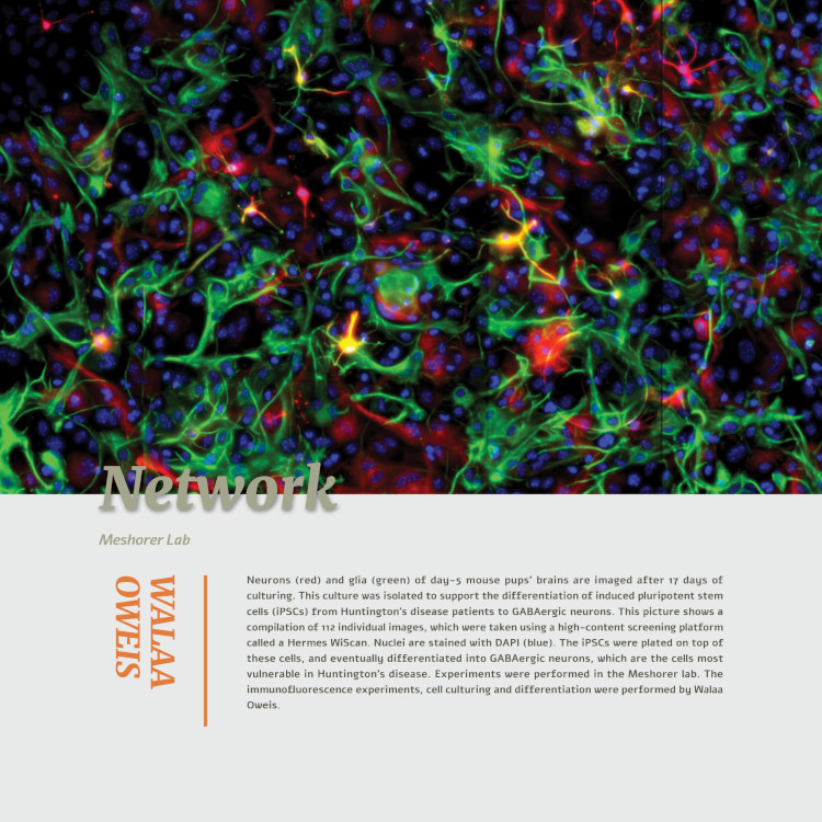
Name of work: Network
Submitted by: Walaa Oweis
Lab: Eran Meshorer
Description: Neurons (Red) and Gila (Green) in a 17 days old mouse’s brain
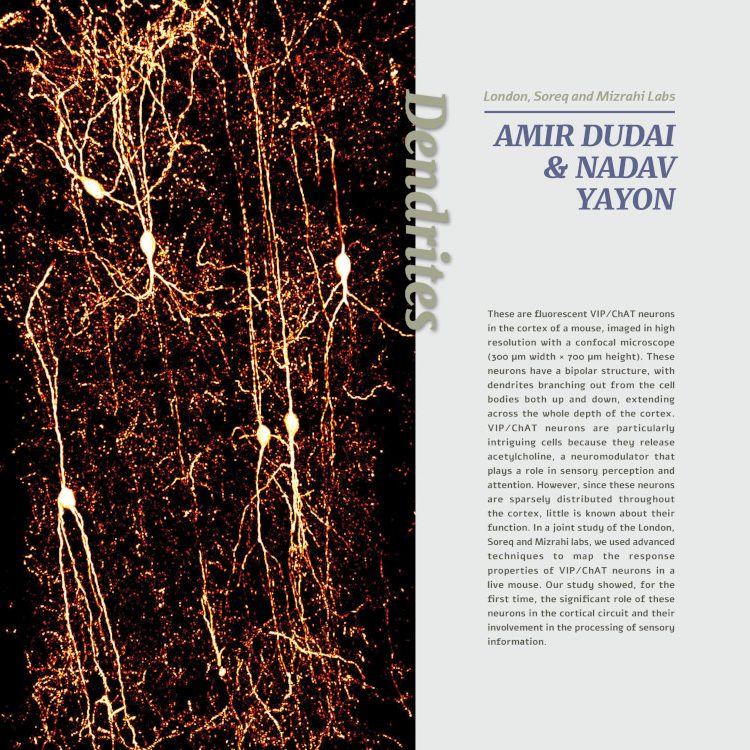
Name of work: Dendrites
Submitted by: Amir Dudai & Nadav Yaron
Lab: London’s, Soreq’s and Mizrahi’s Lab
Description: Fluorescent VIP/ChAT neurons in the cortex of a mouse, imaged in a high resolution with a confocal microscope (300 um width x 700 um height)

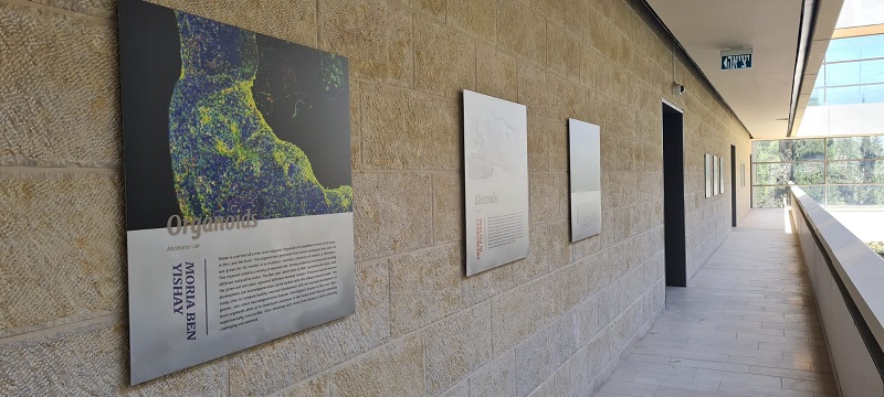
Name of work: Organoids
Submitted by: Moria Ben Yishay
Lab: Meshorer’s Lab
Description: Mini-brain organoid
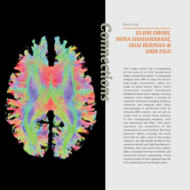
Name of work: Connections
Submitted by: Elior Drori, Rona Shaharabani, Shai Berman, Shir Filo, and Aviv Mezer
Lab: Mezer’s Lab
Description: A tractography of the brain of an ELSC postdoctoral fellow, viewed from above
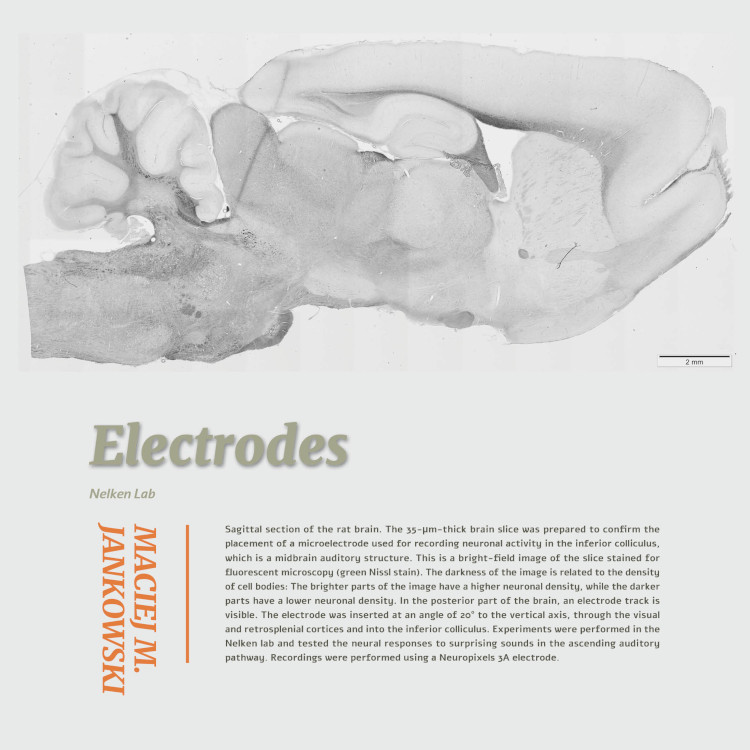
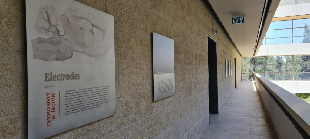
Name of work: Electrodes
Submitted by: Maciej M Jankowski
Lab: Nelken’s Lab
Description: Sagittal section of the rat brain

Name of work: Attentiveness
Submitted by: Adi Kaduri Amichai
Lab: London’s Lab
Description: The locus coeruleus (LC) in the mouse brain

Name of work: Perception
Submitted by: Hadar Levi Aharoni
Lab: Tishby’s Lab
Description: An hourglass-shaped illustration of the brain
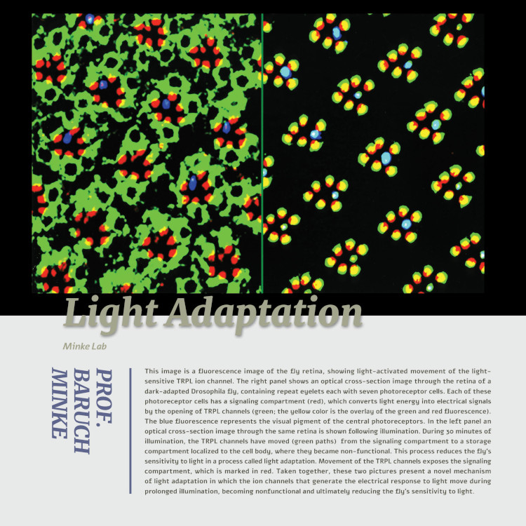
Name of work: Light Adaptation
Submitted by: Prof. Baruch Minke
Lab: Minke’s LabDescription: A fluorescence image of the fly retina, showing light-activated movement of the light-sensitive TRPL ion channel
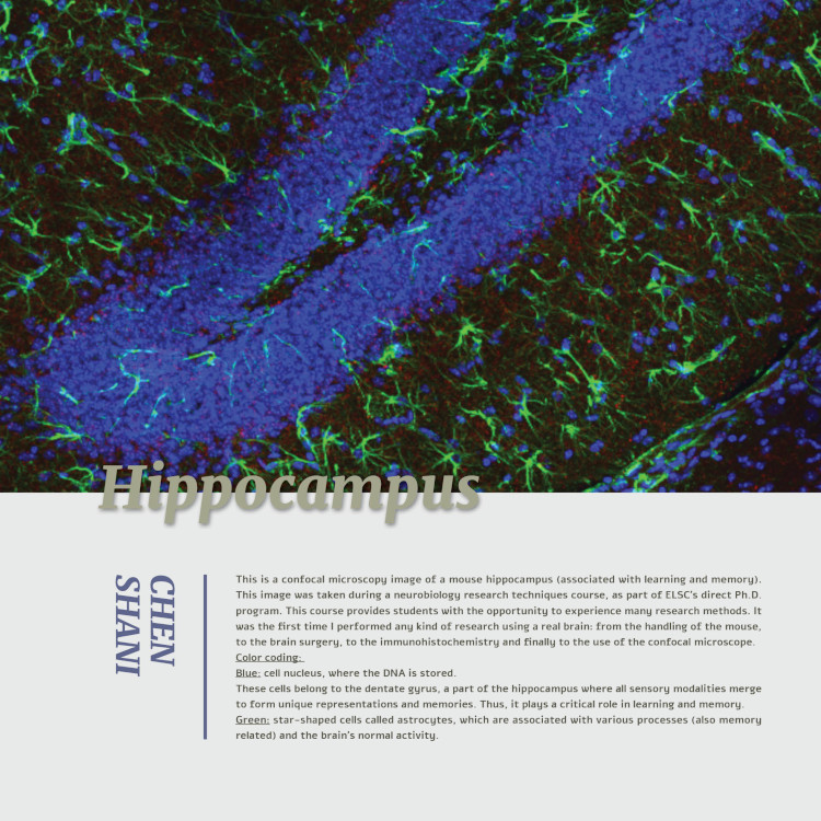
Name of work: Hippocampus
Submitted by: Chen Shani
Lab: Hyadata’s Lab
Description: A confocal microscopy image of a mouse hippocampus

Name of work: Transgene
Submitted by: Hodaya Vrubel
Lab: Habib’s Lab
Description: An image of a primary cell culture containing different cell types from a mouse brain
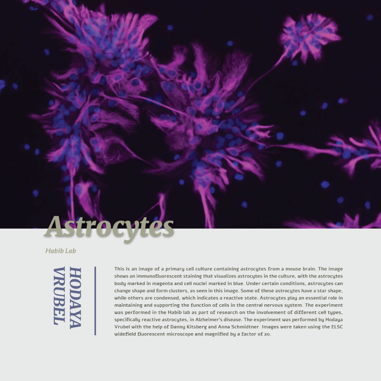
Name of work: Astrocytes
Submitted by: Hodaya Vrubel
Lab: Habib’s Lab
Description: A primary cell culture containing astrocytes from a mouse brain

Name of work: Wiring
Submitted by: Ben Jerry Gonzales
Lab: Citri’s Lab
Description: Coronal sections through a mouse brain
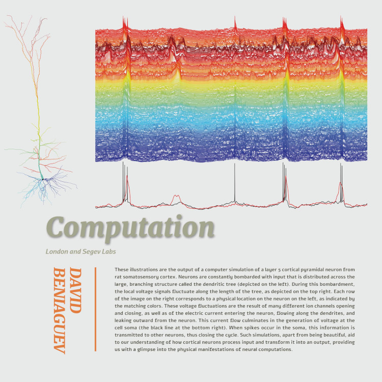
Name of work: Computation
Submitted by: David Beniaguev
Lab: London’s and Segev’s Lab
Description: Illustrations of the output of a computer simulation of a layer 5 cortical pyramidal neuron from rat somatosensory cortex
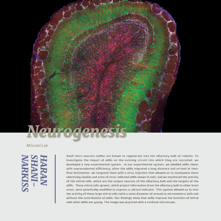
Name of work: Neurogenesis
Submitted by: Haran Shani-Narkiss
Lab: Mizrahi’s Lab
Description: Adult-born neurons

Name of work: Diversity
Submitted by: Or Gold and Adi Ravid
Lab: Habib’s Lab
Description: Cells in the hippocampus of healthy, wild-type mice and of Alzheimer’s disease mice

Name of work: Neuroimaging
Submitted by: Roey Schurr
Lab: Mezer’s Lab
Description: Left and right views of the human brain using MRI