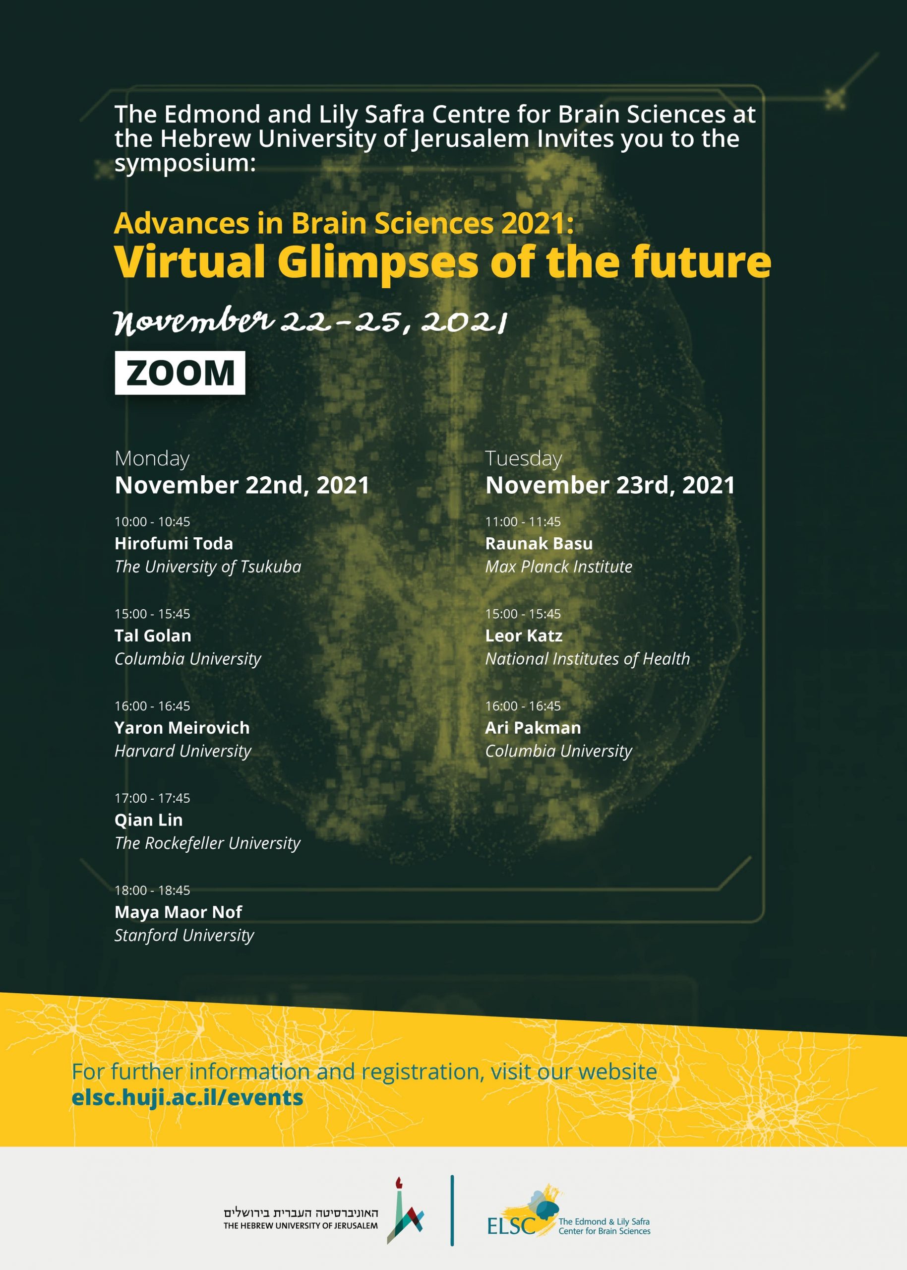
The Edmond and Lily Safra Center for Brain Sciences at the Hebrew University of Jerusalem Invites you to the symposium:
Advances in Brain Sciences 2021:
Virtual Glimpses of the future
November 22-25 2021
On ZOOM
Monday, November 22nd, 2021
10:00-10:45- Hirofumi Toda
The University of Tsukuba
A novel sleep-inducing molecule, Nemuri, links sleep and immunity. Genetic Dissection of Sleep in Drosophila
Sleep remains one of the biggest mysteries in biology. We do not have a satisfactory answer as to why we need to sleep and how sleep is regulated. In order to understand the molecular mechanism of sleep regulation, we conducted an unbiased and genome-wide screen for genetic factor(s) that induce sleep. We identified a novel sleep-inducing gene we named ‘nemuri’, which encodes an anti-microbial peptide. Nemuri is induced under conditions of high sleep need, such as following sleep deprivation and during sickness, and it then drives recovery sleep. Overexpression of Nemuri helps survival of flies after a bacterial infection, likely because increased sleep helps in fighting infection. Thus, Nemuri mediates a two-pronged strategy to facilitate responses to immune challenges—it kills microbes and induces sleep.
15:00-15:45- Tal Golan
Columbia University
Bridging visual object recognition and deep neural network models by means of model-driven experimentation
Deep neural networks (DNNs) provide the leading stimulus-computable model of biological visual object recognition, but their power and flexibility come at a price. Due to their capacity to absorb massive data, distinct DNN models often make very similar predictions when tested on stimuli sampled from their training distribution. To enable continual refinement and improvement of DNNs as scientific hypotheses about biological vision, we must be able to compare alternative models efficiently. I suggest taking a model-driven experimental design approach to achieve this aim. I developed a methodology for synthesizing controversial stimuli: inputs (e.g., images) for which two or more models make conflicting predictions. Empirically measured responses to such stimuli are guaranteed to provide evidence against at least one of the models. Therefore, controversial stimuli allow contrasting the validity of different computational models efficiently. I will present results from experiments employing this approach to compare models of human visual categorization; the translation of this method to the domain of natural language; and the application of stimulus optimization for comparing DNN models of neural representation, using the representation of human faces as a test case. I will outline how stimuli driven by DNN models can form a third category of experimental stimuli in cognitive and systems neuroscience, complementing and bridging parameterized artificial stimuli (e.g., Gabor patches and random-dot stimuli) and complex natural images and videos.
16:00-16:45- Yaron Meirovich
Harvard University
Unraveling the principles of circuit plasticity using Connectomics
Automated large-scale electron-microscopy in the service of connectomics is an emerging field in neuroscience that systemizes the construction of neural circuits at the synapse level. Connectomics, especially when combined with physiological and behavioral assays, opens new avenues to our understanding of neural circuits development and the physical underpinning of learning and memory in healthy and diseased brains.
In my talk, I will present the high-throughput machine learning algorithms and systems I developed for scalable and lower-expense connectomics (PPoPP’16, aRxiv’17, CVPR’19) and their application to several fundamental questions in neurobiology. First among these are complete reconstructions and developmental comparisons of eight C. elegans brains, revealing several new principles of circuit maturation (Nature’21). Second is my work for the last several years to map and better understand the rules that sculpture the neuromuscular connectome from birth to adulthood. We found that the connectomes of neck and finger muscles in mice massively simplify during the first postnatal weeks, and the remaining connections strengthen. Although at birth there is nearly all-to-all connectivity, perinatal motor neurons already co-innervate muscle fibers in a nonrandom way and are associated with each other in a fixed order, perhaps related to their activity patterns. This idea is reinforced by evidence for an activity-dependent elimination rule of entire neuromuscular junctions. Our work provides a mechanism that determines the order of axon pruning at individual endplates, resulting in the global recruitment order among motor neurons. The fact that this coordination also impacts synapses that are 1 millimeter apart on muscle fibers invites speculation about whether such mechanisms might coordinate inputs onto cortical neurons as it does in muscle. Lastly, I will describe my collaboration with the Hochner lab on the construction of the canonical circuit of learning and memory in the octopus vertical lobe. Our data suggests that memory representations are temporally stored in more than 20*106 synapses placed on unique single-input non-spiking interneurons, in a 4-layer feedforward neuronal network, demonstrating the novel network motifs that have evolved in octopuses to subserve learning and memory.
17:00-17:45- Qian Lin
The Rockefeller University
How decisions unfold within the whole brain
Planning of goal-directed behaviors requires the interaction of multiple brain regions, but how brain-wide neurodynamics leads to action is less understood. Moreover, a hallmark of behavior and decision is the variability across trials; this variability cannot be captured by trial-averaged analysis that uses tuning frameworks for behavior representation. However, investigating the neuronal basis of motor planning at the whole-brain, the single-trial level is currently impossible in mammalian brains due to technical limitations. Here we have tackled this question on the whole-brain, single-neuron, and single-trial level using larval zebrafish, by combining high-speed volumetric calcium imaging using light-field microscopy (LFM) with an operant learning paradigm. At the global brain level, we demonstrated via experimental data that decision-making relies on the coordination of brain-wide information – a widely accepted notion but not directly tested by experiments. Within this brain-wide network, the cerebellum displays a particularly strong pre-motor activity, predictive of both the timing and outcome of decisions more than seconds in advance. We developed a quantitative cerebellar model in which the competition of the left and right hemispheric decision signal determines direction while the rate of bi-hemispheric joint ramping activity explains the timing on a trial-by-trial basis. Using cell-type-specific transgenic lines and two-photon imaging, we identified that the cerebellar granule cells are the main population contributing to the observed ramping signal, not the Purkinje cells. Our work is one of the first to apply LFM to neuroscience research, and our results highlighted a cognitive cerebellum, and demonstrated brain-wide information coordination during decision making.
18:00-18:45- Maya Maor Nof
Stanford University
Mechanism of axonal degeneration and neuronal elimination during the dysfunction of neural circuits.
Aberrations in proteins folding, processing and degradation are common features of neurodegenerative diseases, resulting in their accumulation. Amyotrophic lateral sclerosis (ALS) and frontotemporal dementia (FTD) are two neurodegenerative diseases that share genetic and neuropathological features. The most common genetic cause of both ALS and FTD is a hexanucleotide repeat expansion in the C9orf72 gene. In both diseases aggregation of the protein TDP-43 is the major pathological hallmark. How the dipeptide repeat protein poly(proline-arginine) translated from the C9orf72 hexanucleotide repeat expansion and the TDP-43 protein accumulations contribute to neurodegeneration is largely unknown. To study the cellular mechanisms driving neurodegeneration, I developed a platform to interrogate the chromatin accessibility landscape and transcriptional program within neurons in response to pathogenic protein accumulation. I provide evidence that neurons expressing the dipeptide repeat protein poly(proline-arginine), activate a highly specific transcriptional program, exemplified by a single transcription factor p53. Ablating p53 in mice completely rescued neurons from cell death and axonal degeneration and markedly increased survival in a C9orf72 mouse model. Furthermore, p53 reduction was sufficient to rescue C9orf72 ALS patient iPSC-derived motor neurons from degeneration. Mechanistically, p53 is stabilized, binds to DNA and activates a downstream transcriptional program, including Puma, which drives neurodegeneration. These data demonstrate neurodegenerative mechanism are dynamically regulated through transcription factor binding events controlling gene expression programs and provide a framework to apply chromatin accessibility and transcription program profiles to neurodegeneration.
Tuesday, November 23rd, 2021
11:00-11:45- Raunak Basu
Max Planck Institute
Representation of navigational goals in the orbitofrontal cortex
Planning a journey to a goal destination requires representation of the future goal, knowledge about the current location, and a spatial map where the relative geometry between the current and future location is preserved. While neurons in the hippocampus and parahippocampal regions provide estimates of the animal’s current position and nearby trajectories, a specific neural code of the remote navigational goals remains to be identified. We hypothesized that the future goal location might be represented in a brain region outside the hippocampal-entorhinal system, like the orbitofrontal cortex (OFC), which has been implicated in representing decisions in non-spatial tasks. To test our hypothesis, we designed a linear track with multiple reward sites, in which rats were trained to alternate between two given sites to obtain rewards. After few successful alternations, the reward sites are changed to new locations, thereby ensuring continuous update of goal representation. As rats performed this task, we simultaneously recorded the activity of hundreds of OFC neurons. Analysis of the neural activity revealed that OFC neural ensembles exhibit distinct firing patterns at the different reward locations in the maze, thereby encoding the animal’s positions during reward consumption. However, after reward consumption and just before the onset of navigation, these neurons exhibit a representational transition from the animal’s current position to the next goal, which is persistently and dynamically maintained during the entire journey until the animal reaches the destination. Finally, optogenetic perturbation of OFC neurons, specifically at the navigation onset, caused animals to navigate to an incorrect destination, pointing to OFC as part of the brain’s internal map representing the animal’s decision of navigational goals.
15:00-15:45- Leor Katz
National Institutes of Health
Neuronal mechanisms for decisions, attention and working memory in the primate visual system.
Making accurate decisions, selectively attending to certain items over others, or manipulating information in working memory are fundamental behaviors that rely on specific neural circuitry. Throughout my research I have contributed to understanding such behaviors in human and in nonhuman primates but found that despite tremendous advances in the field, we still lack a mechanistic understanding of what goes wrong in conditions such as dementia or autism. My long-term research goal is to determine the neural underpinnings of cognitive behavior, in health and disease.
In this talk, I will present my main contributions towards uncovering neuronal mechanisms for cognition in the macaque, an animal model whose neural circuitry is remarkably similar to ours, has an elaborate behavioral repertoire, and affords an understanding of human brain functions. First, I will demonstrate the utility of rigorous psychophysical frameworks in determining the causal contribution of key brain regions to behavior in a perceptual decision-making task. Next, I will describe how causal manipulations of specific brain structures can be used to identify hitherto unknown brain regions and reveal new functional circuits in support of cognitive functions such as selective attention and object recognition. Finally, I will present my future research directions and approach, which leverage my experience studying how we select from external information (sensory signals) to investigate how we select from internal information, such as information stored in visual working memory. By blending theory-driven experiments with large-scale electrophysiological recording and causal manipulations in behaving macaques, I aim to uncover how we select information from working memory and equally important, how we fail to do so when struck by disorders of executive or memory function.
16:00-16:45- Ari Pakman
Columbia University
Quantitative Tools for Neuroscience Questions
As bigger neuroscience datasets are generated with novel observation modalities, so grows the need for computational tools to answer basic questions. How many different types of neurons exist in a population? How to sort out neurons from their electric activity? How do neurons process information? I will present statistical, machine learning and information-theoretic tools that address such questions. In particular, I will discuss new solutions to the problem of classifying neuron types using genetic markers, amortizing spike-sorting in modern multi-electrode arrays and disentangling the simultaneous presence of synergy and redundancy in neural information processing circuits.


